
04. amen — the two-day workup, july 2026
Baseline: Nov 10, 2025 · Peak Brain · QEEG + IVA-2
Retest: Dec 19, 2025 · Peak Brain · QEEG + IVA-2 · Dr. Hill Zoom readout Dec 31
Amen: Jul 9–13, 2026 · SPECT ×2 + Total Brain + Conners CPT 3 · Dr. Creado
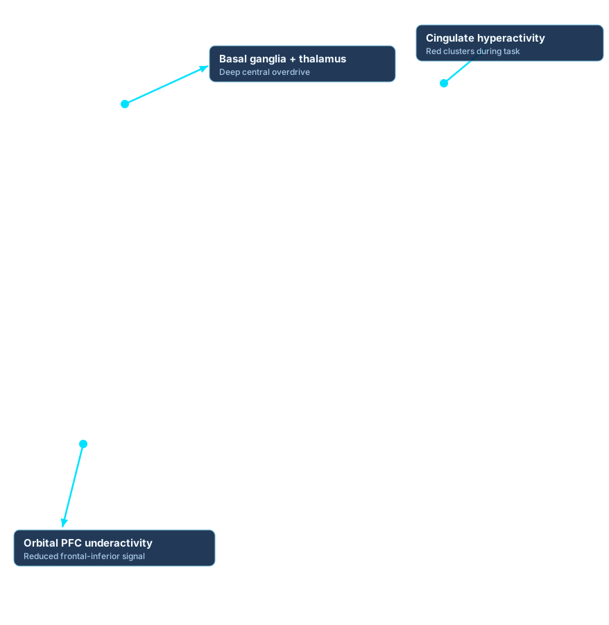
the spect story on one tab: the scans from the two-day workup, dr. shane creado's readout, the total brain assessment, and the amen intake screener battery. the scans below are my own — a concentration-task study on july 9, 2026 and a baseline study on july 10, 2026. each render has a clean view and an annotated view; toggle between them with the control on each card.
on the total brain mental-health screening battery, every indicator came back "at risk: no" — depression (phq-9), anxiety (gad-7), adhd, ptsd (pc-ptsd-5), substance abuse (cage-aid), sleep apnea, and social anxiety.
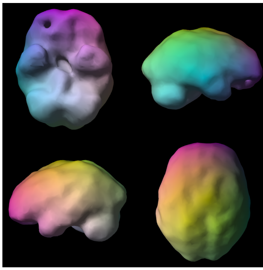
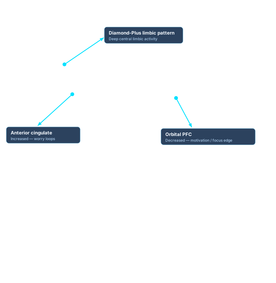
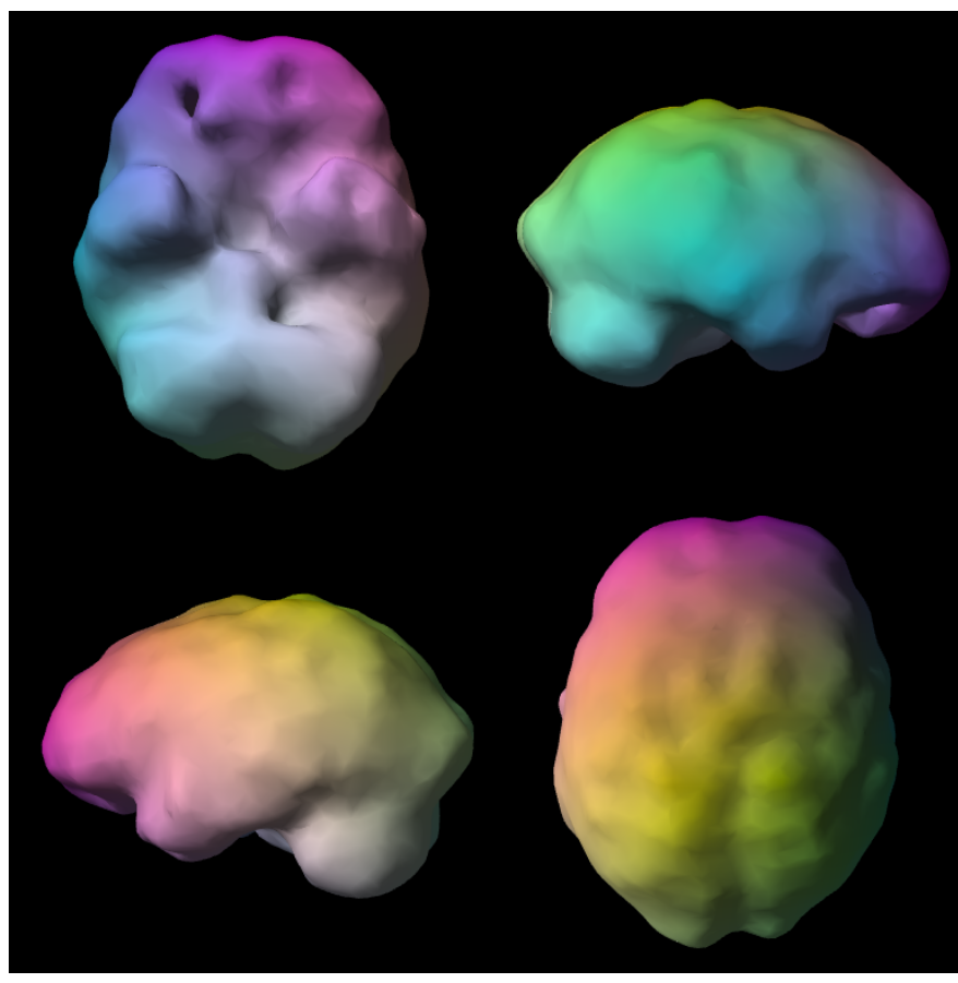
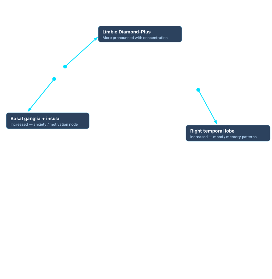
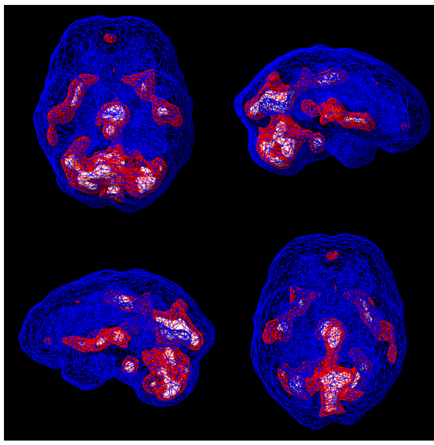
signed july 13, 2026 — shane a. creado, m.d."brain spect imaging is used as an adjunctive source of physiologic information and is not, by itself, diagnostic of any psychiatric disorder. diagnostic impressions and treatment recommendations are based on the totality of available clinical data. brain spect findings should be interpreted within the context of the patient's complete clinical evaluation. imaging findings alone are not intended to establish, confirm, or exclude a psychiatric diagnosis and should not be interpreted as definitive evidence of any specific mental health condition."
that's the ordering physician saying, on the record, that spect is not a diagnostic instrument in isolation. it is the same diagnostic-humility framing i've held through this whole project, now coming from the physician who ordered the scan.
[ 00:04:40 ] — kevin, on the amen visit"with amen clinic, the things that landed with me were just how there was — reflective of the blood flow in the brain — was consistent with some of what they were seeing with the tests. just a little bit of confirmation. but there wasn't any severity of showing severe blood flow changes that were outliers, which i was happy to see. i was hoping that we didn't find anything that was locked or stuck, or whatever words they wanted to use."